
 Serious Inquiries Only
SIO500: A New Study Suggests Brain Imaging Might Have Significant Flaws
Brain pics, or they didn't happen? There's a recent study that demonstrates that some brain region activity might interact differently with oxygen than previously assumed. This could upend significant neuroscience research spanning decades. Thankfully SIO has Dr. Jenessa Seymour to come in and break it all down for us!
We start with a primer on the science of neuroimaging tools and the images they produce. How in the WORLD can we get a picture of a brain while a person is still alive? What are the things we need to know when evaluating scholarly articles or popular media coverage about the brain that uses these brain "scans" in discussing their results? And what do we need to see next to support the findings of this study that could have incredible ramifications across our understanding of the brain?
-
Example fMRI with a more-appropriately blurry sMRI underneath
-
After Warping Options, Configurable Pipeline for the Analysis of Connectomes (0, 4, and 8mm filter images)
-
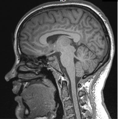
Jenessa's Brain:
Are you an expert in something and want to be on the show? Apply here!
Please support the show on patreon! You get ad free episodes, early episodes, and other bonus content!
 Apple Podcasts
 Spotify
 iHeart
 Support on Patreon
Transcript
This transcript was generated automatically and may contain errors — the audio is the record.
0:00:11 Thomas: Hello and welcome to Serious Inquiries Only. This is, oh my god, it's episode 500. well I didn't know that until now, because I didn't plan on doing a 500 thing. That's pretty freaking cool though. And you know, it works out perfectly because 500 is going to be core SIO. Got a science thingy for you. I especially in this day and age, sometimes it's nice to just learn something, talk about something scientific that has nothing to do with Trump or politics or anything. Just good old science learning. Is a great guest for that. Of course, Dr. Jenessa Seymour. This episode comes from a couple of different things. For one, there's a study that Jenessa references that looks at, well, I guess it's kind of meta. It's not a literal meta study, but it's kinda meta in that it looked at how often studies get cited. And it showed that if there's picture of a brain on the study, as in like an functional MRI, I take it, increases the number of citations that a study gets, which is kind of crazy to think about. And also, I think a lot of science and skeptic minded people will be familiar with the fact that functional MRI pictures of brains, you know, with a little red activity on them can be used to make claims that might not necessarily stand up to scrutiny. Knee. It might be maybe over claiming a little bit. And there's a reason behind that. Something that Jenessa knows very well, from her scientific experience. A, there's always a reason why those functional MRI images can never be as accurate as you might think. And sometimes when you think something's more accurate than it is, that can definitely cause problems. There's another reason we're talking about this, which is there's a recent study that is possibly casting some of this methodology into doubt. And Jenessa will get to it toward the end of the recording, but it's possible that a lot lot of studies that use functional MRI to kind of image where brain activity is happening. It's possible those were wrong based on sort of the science of how that's detected works. A study claiming that as much as like 40 percent of that, might be wrong. She'll tell us more about it, but that would be not great. All this is to say, happy episode 500, happy SIO everybody. Let's learn about brain imaging. Can't wait. Hey, support the show patreon .com slash series pod. More science thingys. Lose the ads and get that warm tingly feeling in your brain we can image. Jenessa is going to explain now after the break.
0:02:43 Thomas: Jenessa Seymour. How are you doing?
0:02:44 Jenessa: Pretty good. Had a nice little white Christmas over here, belatedly, so that was nice.
0:02:51 Thomas: Yeah, no, I don't know what that would be like or what that could be like. No clue. It's never happened. Never going to happen.
0:02:58 Jenessa: I feel like less likely would
0:03:00 Thomas: Where, I'm from, it'S It's maybe statistically happened because it snows like a couple times a year, but not in Sacramento. That's it's almost never. It's like I think I'm at freaking sea level here. Yeah. A good Christmas nonetheless. Are we here to talk about today?
0:03:07 Jenessa: Be concerning. Today I figured we could talk about something that I think people often might not have questioned it, really. But when you think about it, it's a little strange. You read neuroscience papers or when you see it in the news, you know, there's a picture of a brain in there most of the time. Where the hell did that come from? How did it get there? How Did I Get a Picture of Your Brain While You Were Alive?
0:03:33 Thomas: Came my brain. I took a picture of a brain. Yeah.
0:03:41 Jenessa: Like, well, of course, why can't you just do that? But it's actually kind of complicated to do it and to do It Right and to... a lot of assumptions built in to assume that we actually got good information about what your brain was doing at the time. To really be like a critical thinker when you see neuroscience in a scientific article or in the media, it does require knowing a little bit about how we got this. So I we could talk about magnets and how they work and why I can take a picture of your brain with a magnet.
0:04:12 Thomas: Magnets. Finally, we're going to get to the bottom of magnets, everyone. One.
0:04:16 Jenessa: An older paper, but a paper came out that showed you put a picture of a brain in a psych or neuroscience paper, it just gets cited way more often. It gets a lot more attention. And that's not necessarily warranted. There's really good research that looks at how brains work that didn't necessarily take an fMRI specifically of a brain you know um sort of a dazzle me with science kind of effect going on it's
0:04:42 Thomas: And it's already a science thingy on a science thing this is like a meta science thing I have to assume they controlled for everything else and it's like no literally this picture of the brain adds a bunch
0:04:58 Jenessa: Yeah like you could do the same study but you were just focused on response times and like that's a measure of neural functioning like how fast you can respond to something tells me how quickly that signal zapped through your brain and down your arm, or EEG is another perfectly good way to look at signals in the brain, but it doesn't make that pretty picture. So certainly, I'm sure you can imagine, you know, if you're trying to get popular media to put an article together about you, you Know, yeah, if there's a thing that looks like what your average person would recognize as a brain, it just makes for a nice little headline a nice thumbnail and gets a lot more attention but your average neuroscientist can look at that and screw up and not know what the hell they're looking at it's actually kind of complicated and it's really easy to like manipulate that image if you do it wrong or if suppose you could could be nefarious about it. But yeah, generating that image is not like some, it's not a camera, you know, I don't crack your skull open and just take a picture. It's really complex, this process. So if we understand where it comes from, how it got there, we can be more critical in understanding like, what I looking at? I'm not just looking at like color blobs on top of a picture of a brain. And that means whatever the hell you told me it meant, because I don't know what I'm looking at.
0:06:26 Thomas: You're so you're talking about the thing where it's the sort of like a cross cut of brain and horizontal slice of it kind and there's like different like red little up things versus the things that aren't lit up it's like that okay
0:06:39 Jenessa: Yeah, exactly. That's a classic fMRI type image would be showing in always a change in blood flow is what they're looking at, so how the hell did they make that picture?
0:06:52 Thomas: Are we worried about like how they literally made that picture or like how the blood like the more sciencey part of it oh okay all right interesting interesting
0:06:59 Jenessa: Of both. I would say. So like starting at like square one, like why the heck is a magnet even able to take a picture? That doesn't make a lot of sense. Also really important for thinking about, you there's lots of different tech we could use to look at stuff happening in your brain. Like why are we so partial to these MRIs? Thing is it's super safe. So So you go get exposed to a magnetic field and then walk away. And like, as far as anybody can tell, it doesn't do anything to you. People like with those really strong magnetic fields. I know when I get around it, I get like a little dizzy, but like that's pretty much it. It it doesn'T have any lasting effect. You pregnant people can go Get an MRI. It's really cool. You can see like them and the baby.
0:07:43 Thomas: Hmm because realizing it's not like radiation it's not like x -rays okay it's just magnets so as long as you don't forget about any piercings that you have
0:07:52 Jenessa: So the one safety issue the most important thing is that you do not have any metal on you or in you um most of the time people know about that so like check your pockets take your rings off take your earrings out no bobby pins um is a magnetic field like you've never experienced in your life have you ever had an MRI okay
0:08:12 Thomas: I've had a couple yeah on my knee because they messed up the first one then had to do it again and it's basically just a lot of sitting still
0:08:14 Jenessa: So you know yeah pretty much so you do have to hold super duper still because uh think about it like as if it was a camera like with a really long exposure you somebody was doing a long exposure on a camera and you ran past you'd make like a little blurry ghost um taking super duper fine detailed image but taking forever to do it so if you move at all you just ruined it it um know one of the ways that I thought this was so cool johns hopkins has uh experimental uh with kids what they would do to train them to hold still they had like a fake magnet that they could lay down in and they would play a movie and every time the kid moved the movie would pause so it would like you gotta hold real still or it's gonna turn off off everybody um know for little little kids um know like infants they move too much if if they're really infants you just swaddle them up really tight and then they can't do anything um like toddlers an MRI is basically not indicated unless if it's super important you would anesthetize them obviously generally anesthesia is not a great thing that everybody wants to be doing. So, yeah, it's infancy swaddle them. Young, young kids, you don't do it unless it's absolutely vital. And then, you kids all the way through adults just hold real still. Yeah. Any metal off of your body. And if there's metal in your body, you Know, like an implant or something. Modern implants are almost almost always MRI safe. Obviously, you always want to check first.
0:09:03 Thomas: Oh wow diabolical diabolical yeah yeah I don't want to risk that one
0:09:58 Jenessa: You don't want to find that out once you get in there. There is that giant magnetic field in there where, like, if you were wearing a bobby pin, this is not like, you know, the magnet you stick on your fridge. If that ripped a bobby pin out of your hair and it sticks to the side of the magnet, you are not strong enough to pry that thing off. Like, we have to turn the magnet off off to get stuff out and then turn it back on and the way this was taught to me in like the safety classes was like hey first of all it turns into a projectile that can kill people then if we have to turn that magnet off and turn it Back on it's going to cost us like a million dollars in electricity like this is a super cooled coil that is like the power draw to turn, like once it's on, still it's drawing a ton of power. But once it is on, it's ON. To turn it on and super cool it and do the whole thing is like a power draw that is just black out the whole block. Like it's
0:10:59 Jenessa: Unbelievable.
0:10:58 Thomas: I never thought about that so it's like a massive battery kind of thing oh
0:11:02 Jenessa: So there's two parts of the magnet. One is just on and it's generating a electromagnetic field so you go in there and you're sensitive to it um you go into the tube you kind of feel like you're spinning a little bit and it's it's got a positive and a negative and it's you know it's aligning everything um then there's a second part that's a radio frequency pulse so if you hear that like tick tick tick happening uh the more active part Yeah, it's really loud. Yeah.
0:11:26 Thomas: That I remember that it's pretty loud yeah yeah
0:11:34 Jenessa: So when just when the passive part of the magnets on really you walk in the room and all you're hearing is kind of like the cool you'll hear the noise of like all the cooling that's going on. But it's otherwise just kind of the electrical hum of, you And then, yeah, when the machine turns on and the radio frequency pulses start, that is loud. That's why they're going to put earplugs on and headphones and it's pretty loud. What that's doing. Got a lot of water in your body. You're like 70 % water or whatever. Parts of you are more water than others. Like you've got all that liquid in your tummy and your cerebrospinal fluid is a lot of liquid. So that's going to be important for us. Um fatty tissues are way less full of water and water is this polar molecule right it's got a positive side and a negative side so if you I put you in this magnetic field it kind of aligns them all normally you know water is just diffusing throughout your body bouncing around whatever you don't like have a magnetic field but if I putting you in one all your water is going to kind of like line up and the positive aligns with positive or positive goes to the negative negative goes to positive when we hit it with a radio frequency pulse basically um of think of it like you know how the earth is like it's on its little axis and it spins a little bit you
0:12:59 Jenessa: Got those those uh you atoms in there they want to just do that ever so gently. And they're all aligned with the magnet. And if we hit it with another magnetic field, we hit with that pulse, you knock them all over out of alignment with the main magnet. And then kind of like a top, they're spinning, trying to rotate back into alignment with the main magnetic. Is emitting energy. You hit them with a bunch of energy, you knocked knocked them over, and then they go, and they rotate back up with the magnets. So each of those
0:13:35 Thomas: Make
0:13:35 Jenessa: Pulses is, in my head, they do. I feel like the sound effects help me.
0:13:35 Thomas: That sound huh Huh? Okay. Sure.
0:13:42 Jenessa: Yeah, it's called precession. It's that the, you know, the things are just rotating a little bit. You hit them with the pulse, you knock them over. They rotate their way back up like a top, and that's emitting energy. How a machine then picks that up and makes a little picture, that is the last step that I cannot help you with because I don't get it. But basically, that's why there's two magnets. One magnet is aligning everything in your body. And then that pulse, that tick, tick, tick is knocking things back out of alignment.
0:14:14 Thomas: Inside us all, there are two magnets. Got it.
0:14:20 Jenessa: We have added multiple magnets to the situation. What does that mean? We're going to get a lot of this happening in the very watery parts and not a lot Of this happening and the very fatty parts. Your gut, in your cerebrospinal fluid, just a lot a liquid and saline. Gonna have a lot f this magnetic property. And in your very fatty tissue, like body fat, like your axons of your neurons, which are insulated with the myelin sheath to help with conduction of signals, they're wrapped in this fat, that will produce less of this signal. And so that's where you get that black and white image that you've seen any MRI, not just a brain, MRI you've seen with the black and white kind of contrast. Generally, the black areas are more water, the whiter areas are More Fatty. Always have to start there. I'm going to need a structural picture of your brain to start with so I can see like where the pieces are.
0:15:30 Thomas: All right, I'll send you one, I guess. Text it.
0:15:32 Jenessa: Can be extremely detailed, and it really cool to look at. I mean, you don't have to be an expert, like somebody who's been looking at tons of these images, to see that, like, every brain is unique. My heart looks wildly different than yours, like one of us is sick. But brains like to the naked eye, these structural MRIs will, you'll be like, oh, wow, this person's got a bigger this. And this person is like, why is their brainstem so straight? And this one, this thing's kind of crooked. They really are so individual. It's really cool. Definitely if there's something obviously super wrong, you can also like, you do not need need a trained eye to be like, what's that big black spot? That's not, it doesn't seem like it's supposed to be there. So MRI can do a couple of things. Obviously there's the medical purpose. There If will be a tumor that does not look like everything else. Missing piece would be pretty obvious. Even to the untrained eye, you'd be like oh look there's something inside of my head. That doesn't look like it's supposed to be there. There's medical value in that. For the scientific value, you can use this to look at the density of different areas. So how much gray matter do you have? That's the cell bodies. It's got less of the fat. Much white matter doyou have ?That's the axons. It has a lot of that fatty tissue and it looks really white on the images um also some other structural stuff you can do that is like it gets less um in the media but like god it's so cool looking I'm gonna send you one
0:17:14 Thomas: Okay. Love it because we always do this in the group chat with Lydia, and I have to imagine she's
0:17:19 Jenessa: She's just like what the fuck I wonder
0:17:19 Thomas: Just like what the fuck is happening uh yeah, that looks like some finger painting that my kids did. Yeah. Kind of cool though. That that were a painting somewhere in a museum, I'd be like, that's pretty badass. Yes, I like that. It's like, I could tell it's the brain, but it's definitely finger paint that my kids did, better, but but like more organized. Kind of
0:17:39 Jenessa: So yeah yeah Yeah. So, like, these are some of my favorites. This is diffusion tensor imaging or DTI.
0:17:49 Thomas: Is a real image you're saying?
0:17:51 Jenessa: Is a real brain.
0:17:52 Thomas: A real brain. Wow. I love that.
0:17:55 Jenessa: So what they do here is they look at the diffusion of the water. So if, you know, your skull was full of nothing, the water would just randomly diffuse around. Right. Right. It can't.
0:18:06 Thomas: That's what happens to me.
0:18:08 Jenessa: That's what it feels like some days. Can't diffuse through your skull. It canít diffuse through fatty tissue. So when it moves around in the open areas of your brain, itíll just randomly move. But when it strikes white matter, it will move non -randomly. Itíll It'll move along the tissue instead of just moving through it. So these colors are indicating the strength of the white matter, how dense the white matter is in that area. So what you're seeing is all the connections moving through the brain.
0:18:50 Thomas: You're sayings that like on the speed it moves, that it can change color here somehow?
0:18:55 Jenessa: The directionality.
0:18:57 Thomas: Yeah, there's the direction, but there's also color. It sounded like you were saying the resistance or the speed at which it would move would be dependent on the white matter density or something like that. And so is
0:19:08 Jenessa: Be the consistency. Consistency so if one molecule of water if we were in the middle of like an empty space one molecule water is moving to the right one's moving to left one's movement up one's moving down if they were all moving in the exact same xyz direction they must have hit a wall and all moved along the wall instead of moving randomly the
0:19:32 Thomas: I got that. But what's the color then?
0:19:34 Jenessa: Color is the extent extent to which that is true so yeah exactly density is a good way yes
0:19:38 Thomas: Density, maybe. Okay. So, it's not like a speed thing. It's like a density thing. Okay.
0:19:43 Jenessa: Yeah exactly so if there was just like one or two little bits of white matter they would kind of bounce around and kind of maybe move past it in it could move between two axons but if there's a really thick tight bundle of axons it's not going to be able to get through it at all. And so almost all the water will move in the same direction. So that's what this is looking at. And you can find rotating images of this.
0:20:07 Thomas: Wait, wait, wait. We have water in our brain? This is, hold on. What's the, what is a brain? Sorry. Step back.
0:20:14 Jenessa: So yeah, yeah, the cerebrospinal fluid, CSF, it's mostly just water that is all around your brain. It's got like saline in it. Um you know oh
0:20:27 Thomas: Guess I always envisioned the brain and the stuff happening in the brain as being like electrical impulses.
0:20:33 Jenessa: Yeah yeah yeah no the stuff that matters is electrical impulses
0:20:36 Thomas: Okay. But the water is not doing the thinking, but tracing the structure. It's a way to trace the structure for Okay. Interesting.
0:20:45 Jenessa: For purposes of the photo they've taken advantage of the fact that water won't pass through the fat and they're like oh I bet if that's true that will mean the water is going to move non randomly next to the fat right and that's how they get the picture but yeah the way the water water's moving doesn't affect your thought process
0:21:01 Thomas: So cool. Okay, okay, so we can swish that water all around and we're not messing with our thoughts.
0:21:08 Jenessa: It actually protects you from concussions the water so like um you had a brain that's just in air when you like smash your head on something your brain would just smash on the front of your skull um but the water kind of sloshes around with it and gives it a little you know not perfect try not to hit your head but
0:21:27 Thomas: I'm going to do it right now. Let me see. Hold on. I
0:21:42 Jenessa: You can get these detailed structural images mostly useful for medical things but we could also do density um can gets these kind of structural images there's a really cool one one where um test the like tensile strength like how strong is that white matter they lay you in the mri and then they just shake the shit out of you and like the more jelly parts of your brain will kind of shake more and the stiffer parts ofyour brain won't I don't quite know why that matters
0:22:08 Thomas: I can't believe they're allowed to do this stuff.
0:22:10 Jenessa: You know we get away with some weird stuff um forget what that one is called I don't understand what that one's really up to pretty much um I
0:22:17 Thomas: It's called the, Hey, watch this. Let's see if we could get this idiot to just sit here while we shake him a lot. Holy shit. He's doing it. That's what it's called. It's called all of that.
0:22:22 Jenessa: Had much I had friend who doing it in grad school but I couldn't tell you why I who knows um is the one I was telling you about of somebody gave it's
0:22:33 Thomas: I this picture though. Truth be told, it's a really cool picture. Yeah. I really like it.
0:22:39 Jenessa: Beautiful isn't it this one someone giving birth in an mri I don't know why they agreed that's
0:22:45 Thomas: That's not a brain at all.
0:22:47 Jenessa: A brain That's a baby's brain on in a they
0:22:49 Thomas: You're right, babies. I forgot that they do tend to have brains.
0:22:53 Jenessa: Come out with a brainthat pretty much looks like an adult
0:22:55 Thomas: My God. That's always so freakish. I'm sure everyone's seen that meme that's like, here's why they don't do, you why they do the imaging the way they do. Yeah, exactly. Cause the eyes look insane. Like, cause you could see the whole eyeball and it's actually crazy. So the, in this image we can see there's one profile shot where we can can see that and the baby brain wow that baby brain looks like a brain you know which is like not necessarily going be true of everything of a baby like there's a lot of stuff where it's like it's I mean they have a freaking tail and and weird hair you know that brain looks like a brand that's interesting that looks pretty much like what you would recognize it that's
0:23:00 Jenessa: Mris of babies that is terrifying yeah you can even see the amniotic sac bulging at the bottom them it right yeah yep yeah right in like the couple of weeks before birth their brain looks you know it's got stuff left to do on the microscopic level but on a macroscopic level it looks like an adult brain pretty much it's there yeah and then uh I don't know how they got this person to agree to this but yeah they had the baby in there so you see the baby in there and then you see
0:23:46 Thomas: Crazy crazy. Would do that if I were a birthing person. That would be kind of cool. I don't know. They're scientists themselves. Who knows?
0:23:58 Jenessa: I has to be right because like who the hell else would do this I mean its loud its cold this
0:24:06 Thomas: A lot of the best experiments are like, well, no one else will do this, but I'll do it.
0:24:10 Jenessa: Has to be baby number three nobody did this for their first baby
0:24:13 Thomas: I would hope so. That first one, you're afraid of everything. You're just worried about everything. Baby number three, you're like, oh, let's an airplane. Let me jump out of the airplane. And I'll tell you what, But when the parachute hits, the baby will come out and then the baby will have a parachute. They'll have to launch a parachute, too, the or umbilical cord. This is getting gruesome. Okay, never mind.
0:24:22 Jenessa: Be centrifugal force yeah
0:24:35 Thomas: Not that one, but everything else we said.
0:24:35 Jenessa: The the larger point here being it's totally safe baby's fine because
0:24:40 Thomas: Because magnets. Well, that's what's interesting. So like this. I think there's a nervousness around stuff like this, like I think a lot of people if you just told them and you know on one level it's ignorance it's also like I don't know I think it's pretty sensible to be a little bit worried about this stuff because like I think there was a time when you know people didn't know about radiation and how bad that was you know for a minute there and it's like stuff that seems fine all of a sudden can be gruesomely not fine or like down the line cancer whatever but we're confident that just this just magnets just purely magnets there's no like long-term anything there's no it just just of
0:24:42 Jenessa: Magnets yes sure yeah it's just it seems like it if you got metal in or around you you it's super dangerous. If it's ferrous metal, non -ferrous metals will be fine.
0:25:23 Thomas: Course yeah metal what's that okay
0:25:28 Jenessa: F -E -R -R O -U -S, ferrous. Those are the ones that react to magnets. So there's non - ferrous metals that will not react. So you can have metal implants that are non -ferrous. They're designed to be MRI safe and you go in the magnet and it doesn't matter.
0:25:42 Thomas: I got potentially a dumb question what about just like the metal that's in our like body naturally like iron in our blood or whatever the hell like are there any are there with that like is that magnetic at all or does it do anything oh
0:25:55 Jenessa: That's why an fMRI is going to work. Work not in terms of like health like it's not going to like suck all the blood into your face or something um that is what it's going to do it's gonna rely on the magnetic properties of oxy and deoxyhemoglobin to detect changes in your blood flow that's
0:26:03 Thomas: You're right. Yeah. Oh, okay. Oh. I don't know why I was on them. I thought you started with like the water or something. It was like lining up water and all that. I wasn't sure. Maybe I misunderstood.
0:26:19 Jenessa: For the structural so just to get the black and white structural picture of your brain that is a SMRI, structural MRI. The DTI is very colorful that I showed you, but it's still a structural image. It's just telling you where the white matter is. The one where they shake the hell out of your brain, it's a structural thing. It is trying to tell how tense the structural material material is. fMRI functional is telling you what it's actually doing in there. So like on a structural image, I might have a part of my brain that's bigger or smaller than yours, but that doesn't tell me what it is doing, you know?
0:27:02 Jenessa: I could be dead and you could get that same image. fMri, the functional, what we're going to do is we're gonna lay on top of that structural image image, a second image that measured the blood flow. So I'll give you one here. This one's just my brain. Got one done.
0:27:01 Thomas: Right. Yeah, oh, really? You got one for you?
0:27:21 Jenessa: I got a little side shot.
0:27:22 Thomas: Man. I want to see my brain.
0:27:23 Jenessa: Pretty cool, right?
0:27:24 Thomas: That's so you. Not really, but it's wow. You have a very okay.
0:27:26 Jenessa: Can tell, right ? It's totally fine.
0:27:35 Thomas: I You have the, uh, Squidward chin. Don't know why I don't apologies for commenting on anything like that, but it is alarming. I don' it's like, you are the super hot Squidward chin, but I've met you and I don't remember you looking like the super hard Squidward meme, no offense. Uh, so I must be something about MRIs. Like it must be somebody about how it lights up, like certain, like the bone, but not the skin or something like it's, It's not showing certain parts.
0:28:02 Jenessa: It hair isn't there and I'm holding my head in a very specific position.
0:28:05 Thomas: Your goatee isn't there. Exactly. It normally covers it.
0:28:10 Jenessa: Person if I like pull my hair back and like hold my head a certain way you'd be like oh yeah I'll see it
0:28:16 Thomas: Okay. Well, anyway, I think this Hot Squidward is delightful. Full and your your brain looks really cool what's that thing in the center of all our brains that does look like some sort of void there what is that oh
0:28:27 Jenessa: Is one of the ventricles so there is a hollow spot that it's black because because it's full of liquid. It's full of the cerebrospinal fluid. So in each hemisphere, you got a big hollow spot. Like, if you dug down into the center of any one of the sides, you'd find that hollow spot full of water. And then you got two more ventricles as you're going down. See how, like, the cerebellum is that part hanging off the back by, like a tiny little stick. It, there's like a nice little triangle. That is, like the the fourth ventricle, I want to say, third ventricles.
0:29:01 Thomas: I ventricles are heart things too right or am I just stupid
0:29:06 Jenessa: Right. That term is used in the heart, but in the brain, it's a completely different thing.
0:29:09 Thomas: It different oh come on damn it okay it's not just like a generic term for like a of sort void, something.
0:29:12 Jenessa: Know. I are both open liquidy voids, but the heart is doing a lot of active stuff, moving blood around. Cerebral spinal fluid does, kind of flows, but there's not like a pump moving around you know
0:29:17 Thomas: We go. Yeah. Like that. Your The tone there was like, this was one of your children you're embarrassed about. Like, they like yeah, haven't gotten a job yet. It's, it does some stuff, you know, it'S got, it'S GOT a podcast, that ventricle. It'S got it'S, you KNOW.
0:29:36 Jenessa: No no this is um it mean I'm sitting here thinking to myself I'm just now realizing it is the same exact word so it must it must have some sort of latin root that's like just giant gaping maw
0:29:51 Thomas: This like the time cervix and cervical and, the hell out of me. And I to say it on dad's because I was like, hey, I've been too scared to say there's like your spine or whatever. And then there's a cervix.
0:30:03 Jenessa: Yeah are they the same I
0:30:04 Thomas: I don't think so.
0:30:07 Jenessa: Mean well I know it's not literally but it's does it have some latin route I don't know
0:30:09 Thomas: I thought I was coming to you. I don'T know. I still don't know. I looked it up and I the saw answer. I was, like, oh, and then it left my brain because I'm an idiot. So I'll never know. Now, for me, if someone says they have a cervical related thing, it could be that they broke their neck or they have cervical cancer. I don't know. I don '—no, I'm not sure. But I like that you do brain stuff. Like, notoriously, you do a lot of brain stuff, and I was like, yeah, isn't ventricle a hard thing? You're like, never thought about it before in my life.
0:30:13 Jenessa: All right right?
0:30:38 Thomas: First time.
0:30:37 Jenessa: Crossed my mind. Cervical spine. You can see it on this image. It is millimeters from my brain. Couldn't tell you. Why is it called that? Doesn't matter. Irrelevant to me.
0:30:45 Thomas: Anyway, how did we get on this? This is a beautiful picture of brain. I wish I had a picture of my brain like this. It's kind of cool. Yeah
0:30:50 Jenessa: Is cool. It's really cool. So this is all the structural stuff, right? You can get all kinds of interesting structural information about my brain looking at this. This doesn't tell tell you how it's working or how well it's working.
0:31:03 Thomas: Where the thinking happens so
0:31:05 Jenessa: You can pretty much tell that it's, as far as we can tell, it's a physically healthy brain. Nothing looks like it's too bad here to my, you know, not a medical doctor eye.
0:31:14 Thomas: Very very scientific yeah I don't see a lot of badness in the brain I mean
0:31:19 Jenessa: Like missing. Nothing's like dead. It's just kind of there.
0:31:23 Thomas: Again ignorant I would have been like whoa she has a hole in the middle of her brain but I do think that's like totally normal so I'm
0:31:29 Jenessa: That one's supposed to be there. And
0:31:31 Thomas: Assuming since I'm talking to you that's normal but
0:31:33 Jenessa: No holes that aren't supposed to be there so
0:31:34 Thomas: There have been times where people discovered they had like missing parts parts of their brain, right? I mean, it's like, oh shit. And the brain is so plastic that like, you on when and how it happened, you can just be totally normal in all other respects, but you're just missing a big chunk of brain, which is so cool.
0:31:50 Jenessa: Like if that happened to me all of a sudden right now I would definitely notice um if I was just like born missing a piece absolutely like grown adults go get a scan and then somebody's like did you know that you like don't have like almost a whole hemisphere and they're like what are you talking about and it'll just fill in with a bunch of cerebrospinal fluid and doesn't really matter you end up being all right um pretty wild we if people are getting like a ton of seizures in one hemisphere the last resource is a hemispherectomy you take out an entire hemisphere of the brain and if you do it too you would never do this to an adult if you do it to a little kid they like fully recover and they just live a normal life life
0:32:31 Thomas: Many of them become the subject of every philosophy question question ever. Like I took a couple of philosophy courses, which I love, but it's always like, all right, so there's a dude whose brain was split in half. And you're like, oh man, this is like most of the semester was like a dude's brain was spread in half, where you bury the survivors? Like it was that kind of thing, and it's very interesting. Anyway, let get you you off topic. What, what, how do we get back to your agenda here?
0:32:57 Jenessa: So now how do we know what the brain is doing um this is what it would look like if we took that structural image and we pasted on top of it some blood flow see god it is isn't it god damn it
0:33:07 Thomas: The electoral college. Oh, okay. It's the red, red and blue areas and, uh, brain matter doesn't vote. Okay. So it doesn't matter that it looked, the red area looks bigger.
0:33:18 Jenessa: It kind of does in its own way yeah
0:33:21 Thomas: True. The brain doesn't have the stupid electoral college, only we are dumb enough to have that the
0:33:26 Jenessa: You get one vote and your whole brain has to agree eventually something that's kind of cool you can see in the top left see that little white dot sticking off the skull um
0:33:34 Thomas: Oh you mean it's like gray the gray thing like outside of the brain okay
0:33:38 Jenessa: Yeah so the way we make sure that we don't accidentally reverse left and right is we just stick a vitamin e pill like just tape it to the side of your head and uh then I'm like oh I always put it on the left side and that's the left hand of your hand
0:33:46 Thomas: Funny I love that I love the combination of high tech and low tech that happens with some of the stuff often where
0:33:56 Jenessa: Incredibly simple and once we know that works like why would would we ever do anything?
0:34:02 Thomas: Why change perfection? Yeah.
0:34:03 Jenessa: It's working. It's so obvious.
0:34:06 Thomas: It'S cheap and working. That's what science needs.
0:34:09 Jenessa: And it's really helpful because there's neuroscientist view, which is where you put the left on the left because it's the left. And then there's doctor view where you Put the left and the right because if you stood at the foot of the patient's bed and held something up, you want it to match. I'm looking at something that's on your right side I want it to be on your right not my right
0:34:33 Thomas: Fair enough. Fair Enough. Thought that was gonna be stupider, but I, you know, all right. That was at least a reason.
0:34:38 Jenessa: A reason it's really annoying because every once in a while there'll be a paper that comes out of a medical college and less of an experimental place and you better look at that damn legend that tells you well the vitamin e pill is mostly for us so that we know what we're doing but the figure legend will tell you that left is on the right and you're like god damn it because you spent the whole paper going but why are their language centers right lighting up on the way it doesn't make any sense why
0:34:49 Thomas: E pill. Oh, Gotcha. Yeah! My God! This This person's entire brain is switched. Call science immediately.
0:35:13 Jenessa: One's talking about this I'm so confused and it's like oh it was doctors damn it um basically what we do here is we took this very detailed structural image and then this very blobby looking blood flow thing it's a little deceptive there because the resolution the pixels you know the level of precision with which we can place where the blood flow happened is not nearly as precise as the structural. The structural imaging I can get extremely precise. I mean, you can see how beautiful these images are. The blood flow one, if we're being like, I you could say a little more honest, I'll show you here. This picture is like more what the structural imaging would look like if it were on the same resolution. It's kind of blurrier.
0:36:11 Thomas: Yes, the 1984 landslide election. That's right. Dukakis never had a chance. Whatever. Sorry. It's all orange. That's like 8 -bit graphics of fire is what it looks like.
0:36:17 Jenessa: Kind of like, sorry. Yes, exactly. So something that's important to keep in mind when you see a beautiful, super detailed, oh my God, black and white brain image, and then layered on top of it is colorful little blobs. The structural image may have way higher resolution than the blood flow. So your first skeptical point to make sure you put on top of this stuff is that it's giving like a false perception of precision
0:36:57 Thomas: Yeah, that's sort of like, yeah, when you're doing numbers or dealing with statistics and let's say you're you're estimating something and you're like, I am ninety three point two six five eight nine seven seven blah blah blah certain that Hillary is going to win the election. It's you're not supposed to, as a matter of statistics, show that many decimals because it's like, it's dumb. Like there's no, there's No, you would know this better than me. Obviously it's been a minute and I'm rusty, but it it gives would give people a false sense of how precise your metric is because is. So it's, it is almost sort of like that because you've got an image on one hand, you are saying this is really interesting and useful. I didn't know this, the structure stuff, very high resolution. You're you're sort of able to tell with with pretty detailed certainty where this structure is but when you're looking at like blood flow which we do tend kind to use as a stand -in for like here's the thinking sort of thing that's you telling me like this picture I'm looking at is kind of the level of resolution of that oh
0:37:54 Thomas: Because this is dog shit I mean this is like how you could probably tell me but like how many if if I don't know what the terminology would be but like in in video games it would be like a pixel you know or whatever you'd have like you -bit would be a certain number of pixels on the screen etc etc this one if I had to estimate it looks like it's I have 50 pixels by like 70 pixels like total I don't even know like roughly the brain like across length and and width I don't know if you know that actual number but it's it's not much like it there's there's not many pixels on it but it I
0:38:25 Jenessa: Obviously blurry like to the human naked eye this is obviously a blurred up image and
0:38:31 Thomas: Fine granularity. The pixels are the base unit of difference there. So like and that pixel that is the size of, you know, like I'm saying, I mean, there's not very many of them even in this picture.
0:38:42 Jenessa: We talk about it in voxels, which is a volumetric pixel. So instead of like one by one, it's one by one by right. A standard fMRI image, like looking at your whole brain, how many voxel are you going to get um
0:39:00 Thomas: If you've added of depth one, then it's obviously going to be a lot more than I said. Just just FYI.
0:39:04 Jenessa: Yeah yeah so um study is saying 300 000 voxels um keep in mind it's a whole brain critically
0:39:12 Thomas: And it's three dimensions right that you're talking now since you're doing yeah
0:39:15 Jenessa: Three dimensions and one of those they'll usually be somewhere between it's like a one centimeter cube or a two centimeter cube depending on the resolution you're able to get that is going to contain 630 ,000 neurons so
0:39:31 Thomas: And you're getting is now it's I guess it's not a true pixel and it'S not like you're getting an on off right like you'RE getting because I see shades of I see more than two shades so it sounds like you 'Re getting there'S red there'S orange there'S yellow you'RE getting something more than like a binary on off but still you'RE not getting six hundred 130 ,000 pixels for that, you know, or whatever you said, like you're getting, if I were to guess, there's like four, five, maybe different colors.
0:40:00 Jenessa: So you what you're going to do is you're always going to measure against a baseline so this is a difference from a baseline so
0:40:06 Thomas: Okay. It's about how many, sorry, it's about how many shades of difference You Can Do, right?
0:40:10 Jenessa: Yeah that would would depend on the experiment I think that's more within the control of the researcher um
0:40:15 Thomas: Well, I guess, so what are you measuring? Like, what is the, what does the orange, the red, yellow signify? Tell me that.
0:40:21 Jenessa: Always have to check uh the um legend in that study because they might be looking at above a baseline or below a baseline and what that baseline was but in general we are looking at the hemodynamic response so what the hell is that um I got a graph for you here that's going to try to explain it.
0:40:41 Thomas: Blood moving, basically.
0:40:42 Jenessa: Yes. So, your neurons start firing. Now, a neuron firing is extremely fast. Flow does not happen at that speed. What we're really looking for is you, is presented. I show something in your eyes, I tap your finger, whatever. Initially all your neurons are going to fire and we're going to get a tiny little dip dip in the amount of oxygen in your blood because your neurons will consume oxygen. It's oxygen is energy. Oh my God, oh my God. I'm firing. I need oxygen. Give me. The oxygenation level of your blood will dip. Then what we're really measuring is in response, your body goes, oh my God I need blood.
0:41:24 Jenessa: And your blood vessel dilates and blood rushes into the area. So it's actually the increase in oxygenated blood that you're looking for. Critical thing is when people do red and blue, that's not oxygenated and deoxygenated blood. It's almost always above and below a baseline, which is confusing. If somebody told you this is blood flow in the brain, you'd You'd be like, all right, so that part's got the less blood. And it's like, well, yeah, yeah.
0:41:54 Thomas: Sometimes you look at the blue and you're like, those are dead. There's no blood there. Like as an idiot, that's sometimes the thought I have. Wow. No activity.
0:42:03 Jenessa: So you always have to look at the figure legend and see, you know, because sometimes people do on a scale from white is not different from baseline all the way up through like red is very different from baseline and they won't show you anything below.
0:42:16 Thomas: Question I was asking is sort of the degrees of Like that would be the resolution I'm looking for. So, like, I if don't know what the units are, though, so I don'T know what you're you know what I mean? Like, like precision level of difference in blood oxygen, whatever, can it detect is the question, essentially.
0:42:35 Jenessa: You you know, I don't actually know the answer to that. Fairly certain. So the initial dip, the amount of oxygen that your neurons consume is so minimal, it's not detectable. And that's why this has such a lag in response. It takes about four to eight seconds from the onset of a stimulus until this rush of blood to the area is detectable, so the amount that is consumed by the neuron firing while that would be the much more direct measure you wanted that is um typically what you're getting out of this bold blood oxygen level dependent response that's usually um thing that takes about four seconds to show up so it's a huge disadvantage of mri is you can't look at like if something is like this happens then this than this. We're not going to get a time sequence of events happening here. The units of that are, I don't actually know that part. That's the part the software does for me, or really the tech does for me. Has to be more than just the bit that the neurons consume it has to be that giant dilation um correct
0:43:47 Thomas: I guess the point is it's not not an on off. It It's doesn't look like there's like, well, we've detected a change or not, which would be like a binary essentially thing. It seems from the image that there's at least four or five degrees. But those are also you could also be making decision about that. I mean, if it's if I have no idea what units you are. But if you've got readings anywhere from zero to ten, are you are you saying well here's a different color for every one are you saying here's a different colour for every two so there's five different colours you know like that that was the question but if you don't know no big deal but point is based sounds like the me if I'm wrong one of the why the resolution isn't as good is because we're looking at that like blood response that's kind of delayed you know it's like okay this area is pumping pumping a little more blood and that that's, you blood vessels aren't the size of neurons, right? So like, there's going to be like some amount of, is that part of it?
0:44:40 Jenessa: More because that structural response where we look at water and fat that is just a to get a fine point resolution the change in blood flow it's It's harder to get that fine grained of like, we're going to measure that at like 0 .1 centimeter apart from this other piece.
0:45:03 Thomas: I think I'm saying that's why, or I think I was trying to suggest why that might be, but am I wrong about that?
0:45:09 Jenessa: No, no, no. You are right about that. That like we hit some fundamental limitations. I'm like, it's not like we're gonna invent a better MRI and get better at that.
0:45:15 Thomas: You can't get a better like lens and all of a sudden we can see better. It's like the way that this data is essentially collected. It can't really be than that.
0:45:25 Jenessa: It's looking at the blood vessels and like and the way that blood flow changes over time. And like at some point that can only get so small. That is astronomically bigger than the neuron.
0:45:38 Thomas: My point. It's like not you're delivering blood to each neuron at a level where it's like, oh, we can tell that it's going to this neuron, but not that one, essentially. So that sort of limits us.
0:45:47 Jenessa: Now, granted, the structural MRI is not capable of seeing an individual neuron either, but still way, right.
0:45:54 Thomas: But it was just an example. Yeah.
0:45:56 Jenessa: No, you're exactly right. There's no level of precision where blood flow could ever get to the individual neuron level, not even close, because we do not feed individual neurons with individual capillaries. It does not work like that. So it will always be fundamentally limited in its resolution. And just the way that we measure instead of like the procession of the water thing, doing the like oxygenated and deoxygenated hemoglobin and the difference and the fact that you have to measure it in a shorter time frame. You know, that MRI, you if you've had one, a structural one, you know how long you freaking sat there. I have to present you with a sensory stimulus and then I have to wait four seconds and then like, that's my window. I've got like a couple second window opportunity to get the image. It's just kind of inherently a blurrier image because I can't scan your whole brain over the course of 45 minutes. That's how you got that super detailed image of my brain is like like, 45 minutes of me just holding stock still to get one picture of it, when we go to get that blood flow, I gotta get your whole brain over the course
0:46:02 Thomas: Gotcha. Interesting. Right?
0:47:04 Jenessa: Of two seconds. It's just blurrier.
0:47:04 Thomas: You can't tell the blood. Okay. Blood freeze for the picture.
0:47:07 Jenessa: Exactly. That's one thing to keep in mind, is if you see super duper detailed structural and then this more fuzzy blood on top of it. Yeah, don't imagine to yourself that you you have this precise knowledge, that little blob of red came from that exact structural spot, it might give you a slight misperception of what that really is. Also have to think about multiple comparisons here. Think about how many voxels we just talked about. Should not be be doing just an exploratory look at every single little one by one centimeter cube of brain and not correct for multiple comparisons and just say was there a difference in the blood flow yes is the answer there will be tons of them it should look like a shotgun blast if there's no real effect um could end up with a lot of false effects showing up also going to smooth it out a lot because one of the other things we also know about brains is that the effects don't happen in like random one by one centimeter cubes isn't blood flowing in the ventricles because that aren't neurons there we're going to kind of smooth it and I've got an image for you where um do if you're familiar with uh this will actually probably are familiar with this from audio like smoothing stuff like a gaussian um we take that kind of image and we don't smooth it you'll just have like some random voxels pop up because no matter how much correction you try to do for multiple comparisons um there will always just be some random thing that cross the statistical threshold by
0:48:49 Thomas: Oh, kind of.
0:49:07 Jenessa: Chance and we know it's not real because it's either it's in the ventricle or just like it's just not how brains work that just like one one by one centimeter square activated in response to the stimulus and none of the neurons anywhere around it like no no, actually brains operate on the basis of like particular gyrus, this wrinkle in the brain is responsible for this activity. So it shouldn't just look like a shotgun blast. So we do this filtering to then add like, hey, it should kind of be over here and like not telling it where it it should be, like, it should be in this brain area, but more like, if this one is surrounded by every other voxel anywhere near it is not active, ahead and make the assumption that that was bullshit.
0:49:06 Thomas: Hmm, hmm, hmm. Or it's just so it could be your brain doing a different thing than you're trying to look at like it was maybe responsible for.
0:50:04 Jenessa: Theory, it does mean the blood flow did go up right there, but not not, because of the task at hand. Went up for some other reason. But if all the other voxels around it it also went up right at that exact time, that might mean something. So you apply these filters that are essentially going to say, take the activity that is next to your voxel and kind of average it into you.
0:50:31 Jenessa: And that'll sort of cancel out the ones that are random and magnify the ones that are not. But you can overdo it. So the image I sent you is like no filtering, four millimeters. Millimeters that's normal that's how brains work eight millimeters and it just looks like I made three perfect circles and that's probably not right so you have to think carefully before you make these decisions um it's going to produce things that on the one hand are probably more accurate and on the other hand are not what the data looked like so there have been been done to this picture this is not what just popped up oh
0:51:11 Thomas: That's that's really interesting. And that's all going to be obviously that scientists in good faith are going to be making the best decisions they can. But it's just a place where it's like all kinds of room for, you know, human error or just like, you statistical weirdness to kind of creep in. And I that one's interesting. The zero millimeter, the four millimeter, the the eight millimeter. I don't know what, what does zero, what's zero mean? It's just less than filtered.
0:51:33 Jenessa: So zero means we didn't filter at all a zero millimeter filter none filtering yes
0:51:39 Thomas: It's sort of, it's almost like, I know it's probably not literally this, but sort of like a rolling average in a way. It's like, you're taking a rolling average. If I was looking at, you by weeks or whatever, and I'm like, all right, give me the rolling average for like five weeks or rolling average for one week. And you can kind of use that to smooth. I think it is kind of like that you can use that to like yeah smooth out the data and look at longer trends rather than you know obviously you have the immediate one would be like well there's your daily difference in you just say like downloads or something just because I just to bring it to podcasting or I can look at like well that but that's weird because you have different release days and who knows and whatever so like I can Look at a rolling average by the week I can look at a Rolling Average by the by the five weeks so it's like that and it's still there's There's plenty of room for like, to your point, the biggest one, it's eventually if you go to a full degree, it'S going to smooth out to one thing. Like if you're just like, give me the average
0:52:35 Thomas: Of all the data I have, it'll be one number. Useless, pretty much. Maybe not entirely useless, but it's like pretty much it would just be one color that the brain would be. And there's somewhere in there that you can dial in like the amount that that like, OK, this is showing activity in ways that's meaningful and now are there rules or you just kind of trying to dial it in based on what seems right using, you know, all your kind of analytical tools?
0:52:35 Jenessa: Doesn't make yeah Right. Little bit of both. So there'll be industry standards of like, you what's acceptable and there'll be a range of what's accessible. So like I would look at this and just from years of experience, looking at that four millimeter one, yes, there are still a couple of voxels that are active in the ventricles, which is physically impossible. There was not blood flow in a place where there are no veins that is a statistical artifact some statistical artifact happened in the ways that the data came in you know um probably something from a neighboring area or just like the way the baseline was taken um
0:53:24 Thomas: Well, then how is that that happening then? Huh? We are looking at a cross cut. Right. Right. So that's a question I had, too, is like, are about it being of what was the term you used? It was that was like volumetric kind of. I
0:53:49 Jenessa: So like the zero millimeter we would call these voxels because it's a volumetric pixel once we've started filtering we start calling them resols a resolution element because now it's no longer this beautiful little one by one by two by two two by two cube.
0:53:53 Thomas: Voxels. Yeah, sure. Hmm. If it is three dimensional, I've been wondering, like, does that mean you're getting data for all three dimensions? And then whenever we see these images, just by necessity, we're seeing a crosscut of like what the like what's the crosscut we're seen and why.
0:54:23 Jenessa: Pick in advance where you're going to cross through. So it would all depend on what area of the brain you come in interested in. And that's a good thing because you're not trying to test the whole brain or else you're gonna find something. You know in my dissertation research, I wanted to look a visual cortex. And the best way I'm going to be able to do that is these are called an axial section. So that's like starting at the top of your head. If we were to like cut down like towards the eye, then towards the nose, then towards the mouth. That's a really good way if you put it in the right spot, you could get straight through the middle of primary visual cortex. I wanted to do people's primary auditory, the location of primary auditory actually varies so much from one person to the next. I didn't have a particular slice I was going to look at. I was gonna look for a landmark on each person. There's a very characteristic little bump I was going to go look for and align everybody's brain to that point rather than aligned to like central points. So it all depends on what area you plan to look at. And you should make this plan in advance so that you're not going on a fishing expedition.
0:55:41 Thomas: So you you're not necessarily gathering three dimensional data. It has to be somewhat three dimensional, but you're Not.
0:55:47 Jenessa: Data exists. This so you could always
0:55:50 Thomas: So you are gathering kind of everything. Then you're just just what you're looking at.
0:55:55 Jenessa: That you don't fish is by solemnly swearing not to fish you will always have the ability to analyze the whole brain how
0:56:01 Thomas: Gotcha. Guess what the reason I even started talking about that is, is is that any of the reason for this is that it's picking up something above or below the area where you're actually having the cross cut that we're looking at?
0:56:13 Jenessa: So I would say it is possible that what you're spotting could if you ended up in just the right position could it be that you picked up something that's like it looks like it's coming from here but it's actually like slightly below but again these are like at most two centimeters by two centimeters so it shouldn't mess it up too much um a huge amount of space in neuron speak um terms of a major structure
0:56:39 Thomas: Well, Well, what alarmed me was just when you're saying it's literally impossible in that void area. That seems weird. I
0:56:45 Jenessa: That's more where I think about the fact that um is an organic thing that cannot in fact be restricted to a cube and we made it into a cube. And so just statistical artifacts have occurred. You know what I mean?
0:57:01 Thomas: Gotcha. Yeah.
0:57:02 Jenessa: Limitations of the nature of this data collection. The fact that I am using a magnet to try to take a picture of your squishy parts has inevitably resulted in something that doesn't make sense in a couple of spots. The best way I can explain it without um knowledge of the computer science that went into it um that's why I would say like I look at the zero millimeter and that's obviously way too much scatter to be a brain that can't be right but when I look a four millimeter one I say okay there's a couple of spots that like that that can be right that's not what happened but the rest of it looks pretty pretty good. So you know what I'm not going to do is overfit my model. Would not fuss with this any more than is needed, especially if I came in with hypotheses about, you know, these two spots in the back and one spot in the front. And maybe I didn't have a hypothesis about that third spot in the black, but maybe I'll talk about it. Maybe I won't, but I didn'T have a hypothesis about something weird happening in the center of the brain. I wasn't planning to to talk about it anyway there's a little something there oh shit happens the thing on the right with the eight millimeters it's just too dang smooth that
0:58:17 Jenessa: Also looks not real it like at some point you get enough experience with this where you're like that's that's also giving overfit vibes like it just doesn't feel right um more than just vibes to this like I would go if I was you know doing this study I would go read the papers and see what have other researchers done what filters have they chosen what's appropriate in this context um yeah at some point you you enough of these studies and you read enough these papers that like some of these just jump out as like oh some first year grad student didn't know what they were doing made a picture that's too pretty
0:58:51 Thomas: That's interesting. So I guess my question, and hopefully we've described it enough because it's like the zero millimeter, you know, it's kind of spread out a little bit. Four millimeter ,you're almost seeing these clumps. And eight millimeters, basically circles almost because it is made, again, it is smooth and averaged out. So sure. I almost thought maybe you were going to say the 8mm is about as useful as we get, because guess I would ask then, what can you get from the 4mm that you can't get from 8mm? So is it true that like those little arms that are coming off the circle are meaningful enough that you can say we want that level of detail versus what the eight millimeter, which is just the big circles, tells you is, well, you got activity around here, roughly.
0:59:32 Jenessa: So actually I think what you're saying it makes a lot of sense you could make an argument for both lost precision on the 8mm, but maybe that's in a good way to be able to say, let's not pretend I have precision I don't really have. It
0:59:47 Thomas: That's your decimals again on the percentage chance that Hillary is going to win.
0:59:51 Jenessa: An effect in this general area. You could look at is the one in the back. Back on the zero and the four you can notice there's like it's almost actually got two hot spots it's most noticeable on the four there's Like a one very yellow spot and one very Yellow spot and in the eight when it's averaged it together it's actually made a lot of yellow in the center that wasn't there so that may actually smooth too much in the sense that that the there were two voxels that were very active, and this has decided the way to make that not look crazy was to say that the voxel's in between them were also very active. And maybe that's a little too much. Maybe that's becoming less true. So yeah, you're always kind of looking for like the you filter a bit should be trying to get you closer to like what we know is true about brains and especially about blood flow in brains that it doesn't go to like tiny little isolated spots. It goes to specific gyri and sulci the the parts of the wrinkles but you don't want to do it so much that like if two blobs that were separate merge into one that creates an impression that a gyrus in the middle that actually didn't have activity all of a sudden now it does so like where do you draw that line there's some subjectivity to it there's some industry standard to it. The exact right answer is always going to be a little tough. Actually a recent paper out that is calling some of this into question. So the research that initially looked at this, how do we know where we get that hemodynamic response from? Scans. So that's positron emission tomography. That requires injecting you with radiation. We
1:00:43 Thomas: I didn't notice that that makes sense I right
1:01:51 Jenessa: Do radioactive glucose, for example, and then your neuron consumes the glucose and it kicks out radiation when it does it. And we can see that I am pretty damn sure those neurons were firing more than the other ones. But I also had to inject you with radiation. You do these together, we're able to figure out, and they did the initial studies with a lot of basic sensory Neuroscience, hey, it up. So the area that had a lot of activity when we showed you this visual thing, when I tapped your finger or whatever, that area also had blood oxygenation went up when it fired more than baseline, it went down when it fire less than baseline. A recent paper suggesting that might have held up in sensory areas and it might might be less true in other areas of the brain. So if,
1:01:51 Thomas: What are sensory sorry I don't know oh
1:02:45 Jenessa: Auditory, tactile, the areas that process, yes, yes. Sensory processing. So my research is safe.
1:02:48 Thomas: So actually sensory oh gotcha okay okay right so it's so okay so the paper is suggesting that possibly if we were using this like what kinds of of things things would we someone have been using this for that might be called into question like hey the thinking for this certain thing happens in this area versus hey the brain activity for when you're looking at you know an apple or whatever the hell is in it so there might be a difference there oh
1:03:14 Jenessa: What they did was um had people do a math task and a memory task instead of look at this object listen to this sound and didn't do a PET scan because again it's like getting people to sign up for radioactive stuff is not fun but they did um different form of fMRI analysis that looks more at oxygen consumption compared to this bold blood oxygen response. And they seem to have indicated that up to 40 % of the areas they looked at for math and memory were actually showing the of the response you would would have expected that the area was being used for the task and the more oxygen consumption from this one type of measurement actually the blood oxygen response did the opposite of what you expect it to do so uh -oh if that holds up we may have a big problem for quite a bit
1:04:15 Thomas: What kinds of yeah give us a sense of the scale of what that problem might be
1:04:19 Jenessa: The worst case scenario um needs to go back and reanalyze like like decades of memory research for example that used fmri it could be really bad um know I'm sure people are going to challenge it on like somebody needs to do this with a pet scan to really confirm it someone needs to be willing to sit there and take the radiation sorry everybody
1:04:42 Thomas: Gotta get in the radiation box somebody do it come on very
1:04:45 Jenessa: Yeah I feel pretty confident that the sensory stuff is safe that was you know that's how we we established this, that it's going to hold up. And luckily that's what I do. So I'm going to just keep going on. Yeah. Right.
1:04:57 Thomas: Convenient a little too convenient wow yeah
1:05:03 Jenessa: Something to think about as could turn into a big upset.
1:05:06 Thomas: That would be sad that would mean we a lot of stuff we thought we knew we don't quite know essentially right I
1:05:12 Jenessa: Yeah and because if it was 100 % you would go, oh all right, so just go back to all those papers and just just exactly flip -flop everything but it's if it's 40 of the time then it's like well shit what um
1:05:25 Thomas: Don't think I understand what you mean by that but uh okay
1:05:29 Jenessa: They looked at brain areas that respond you're doing a memory task so the baseline can be hey just hang out in the machine and we'll get a baseline of what your blood flow is sometimes the baseline task is like something other than a memory test you know um there's a math test then it can be baseline is just sit still or baseline is do a language task because that's very different from math or something so we get a baseline of what your blood flow looks like in your brain and then we do the test and we see how the blood flow changes because we know there's always blood flowing in your grain um then certain areas of the brain light up different different and what you would have thought is if they're above baseline then there was more neural firing because more blood went to the area and what they're saying is in up to 40 percent of the areas that activated during the task that showed more blood oxygen level dependent change above baseline, that wasn't more firing. It was less oxygen consumption, which
1:06:35 Thomas: Less what and so
1:06:38 Jenessa: Doesn't make any sense.
1:06:38 Thomas: How do they know that again sorry what test are they using to know that
1:06:41 Jenessa: This is what's weird. Still using the fMRI. There's some other analysis method that I have never used before. It's like OEF, oxygen efficiency something. And it's a way to be more closely tied to the oxygen getting consumed I don't fully understand it and that's why I'm saying I think step one is everybody who does not want their whole livelihood overturned is going to be like no no no do a pet scan my guy yeah
1:07:07 Thomas: My question would be why don't they make the whole plane out of that that kind of like why would if that's a better way of doing what sounds like the same thing you're doing doing why wouldn't that have always been the way is it a new method someone invented or what okay
1:07:24 Jenessa: I gotta be honest with you I not following it I tried it's some sort of quantitative method so like part of me no it's not no no no um it's definitely it from everything I've seen it's older than that like it is something that you know like some sort of mathematical something that a human actually developed um far as I can tell jesus I mean if it's some ai bullshit then I would just throw this right out the window but uh yeah yeah yeah I'm am hopeful that probably what this will come down to is a very nitpicky debate about the exact methodologies that everybody should be using. You it's interesting for sure. This has
1:07:28 Thomas: Is it ai just say ai they used ai okay yeah I think that would be really easy to dismiss yeah yeah yeah
1:08:06 Jenessa: Always been an indirect method compared to something like EEG, where we have detected a change in an electrical field because there is an electrical thing happening in your brain this is always indirect and the time lag is so big like it wouldn't be the most shocking thing in the world to hear that it's not doing the best job of picking up what I wanted to pick up you know what I mean
1:08:31 Thomas: And why can't we do the remind me why can't we do the EEG thing is that just not able to I
1:08:38 Jenessa: No plenty of people still do eeg eeg's great oh
1:08:41 Thomas: Mean for the the science like that you're doing doing there's a reason they're doing the fmri instead of that right
1:08:47 Jenessa: So the best thing the thing MRI is really, really good at is this spatial resolution. Think about the location of where this stuff came from, the beauty of that picture of like, oh my God, look at how fine grained. EEG just doesn't do that. It is not even close on spatial resolution, so trade -off there, EEG has incredible temporal resolution timing when exactly stuff happened, terrible spatial resolution. MRI has unbelievable spatial resolution, especially the structural MRI has extreme spatial resolution but the temporal sucks. So we have trade-offs basically.
1:09:10 Thomas: Gotcha
1:09:31 Jenessa: Can my MRI do? Very detailed structures, tractography, where are my white matter tracts, it can show me changes in blood flow, and it can tie a change in blood flow to a structure. So the function there. It cannot give me good timing. When did stuff happen relative to a stimulus? It cannot take a picture from anybody with metal in their body, and it cannot take images without making a lot of noise, which is unfortunately a problem if half my participants are deaf and half are not.
1:09:30 Thomas: Gotcha oh oh
1:10:06 Jenessa: Separate issue that I always had to deal with so
1:10:08 Thomas: Yeah I didn't think about that all right so I'm hopeful that this doesn't mean that all science is ruined but
1:10:15 Jenessa: No I wouldn't go that far yeah
1:10:17 Thomas: Interesting and we'll have to keep an eye on it because
1:10:19 Jenessa: I definitely didn't want to not mention it that somebody just dropped a bomb yeah
1:10:22 Thomas: Might be looking for a new job now just kidding I guess you're not not doing that work anymore but yeah but still really interesting uh we'll keep an eye on it so I've learned a lot about uh and it sounds like in general I guess overall takeaway about the limits of functional mris um not going to be showing you the level of detail that we think I think they are, I guess.
1:10:46 Jenessa: Yeah so their spatial detail is still really good but it's functional part is less detailed than structural so that can be a little deceptive the temporal is terrible don't let anybody try to tell you oh we know exactly when this happened there's people have tried certain statistical modeling I'm skeptical of it it's can be better than nothing but like it's not going to be on the millisecond level ever it's physically impossible and um the biggest thing is just always read that figure legend because sometimes they do red and blue and sometimes they do light red is below and dark red is above and you're like why would why would you do this to me some of them are only ever showing this image the red means above baseline and in the image right below it the Red means below baseline but significant and it's like some of them have left on the right so you just have to read very very carefully when when you look at these, also
1:11:47 Thomas: Just have Jenessa do it for us, everybody.
1:11:49 Jenessa: An option.
1:11:51 Thomas: All right. Well, that was really interesting. I love a good science thingy and really cool to learn more about that. Send us an email if all of science is ruined, I assume.
1:12:00 Jenessa: I'll keep you in the loop. All
1:12:02 Thomas: All Right. Well Dr. Jenessa Seymour, thank you so much. And thanks everyone for listening.
1:12:06 Jenessa: Good talking to you. Button that's called screenshot should take a motherfucking screenshot annotate
1:12:32 Thomas: Can take a picture of your brain but she can't take a screenshot of her computer everybody what
1:12:39 Jenessa: Cat is throwing up in the background I hope that's not on mic um then they go, and they fact that I am using a magnet to try to take a picture of your squishy parts has
1:12:43 Thomas: Science immediately.